“A stark reminder of the human side of what may often be seen as statistics… The book presents, in its clearest form, the suffering of those left damaged by medical complications. It makes for compelling reading for ethicists, lawyers, and patients.” – Journal of Bioethical Inquiry, November 2014
“I believe this book should be required reading for all resident physicians and health science students” – Dr. David Mayer
“Author Dan Walter offers a unique education from the patient family perspective about the damage that corporate greed does to healthcare in general, and the doctor-patient relationship in particular.”
– Student National Medical Association Journal 2014
– John T. James, PhD, Patient Safety America
∞
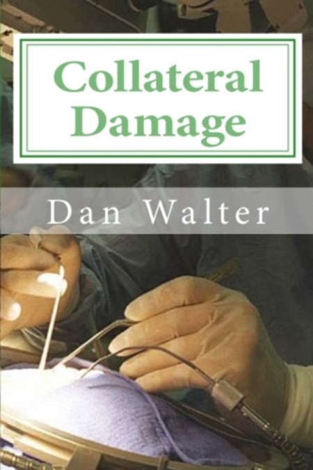
Collateral Damage: A Patient, a New Procedure and the Learning Curve
Preview CHAPTER ONE:
“A Mitral Valve, Flapping in the Breeze, Prolapsed into the Atrium …”
JOHNS HOPKINS MEDICINE has a long tradition of prioritizing patients, and striving for the bottom rung are the anonymous poor. If, for example, you catch a bullet on a Baltimore street corner, or your mother presents you at the ER as a feverish welfare child, then it’s open season for the med students, well meaning as they may be. They can practice on you because if their actions result in an adverse outcome—which is to say that if you are mangled or killed—nobody will question said outcome, precisely because… you are a nobody.
At the other end of the spectrum are wealthy and prominent patients, who get treated by doctors who have already learned what not to do from the mistakes inflicted upon the lower classes.
My wife landed somewhere in the middle. We got snookered by all the hype from US News into thinking that she was going to be treated by the best doctor at “The Best Hospital in America.” Hugh Calkins, MD was to maneuver tiny wires around in my wife’s heart and burn scar tissue in the wall of the atrium to stop atrial fibrillation.
The job required someone with a cool head and a keen eye, and Hugh Grosvenor Calkins, MD, FACC, FAHA, FHRS, Professor of Medicine, Director of the Electrophysiology Lab at Johns Hopkins University School of Medicine—and graduate of Harvard Medical School—assured us that he had done plenty of these procedures, and, he said, “experience counts.” So we knew we were in the best of hands. What we didn’t know is that Professor Calkins—according to what he later told colleagues—follows the practice at most teaching hospitals wherein “the attending shows up to be there during the burn.”
What he meant by that was this:
The patient is etherised upon a table and wheeled into the lab. A student of the treatment performs routine aspects of the procedure. According to the rules, all this is to be done under close supervision. Under bright white light, the rookie’s blue latex fingertips feel their way up the smooth soft skin of the exposed groin, goose bumps because it’s cold in the room, pressing and probing and picking a spot, the point of a large bore needle pressing into the skin until it punctures and there is blood.
The trainee then inserts the sheath for the catheter into the vein and snakes it up into the pumping heart. The catheter wire is then fed into the sheath. This is the point at which you would expect the experienced attending physician to step in because it is a very tricky business to navigate a thin wire around in a beating heart guided by cloudy X-Ray imagery, even if you know what you’re doing.
But since he only “shows up to be there during the burn,” Hugh Calkins was presumably relaxing with colleagues down in the doctor’s lounge or out selling TASER guns while a young cardiology trainee by the name of Richard Wu—whom we’d never  met—was sweating out a decision in the lab. He had a stranger laid out before him and a new type of catheter in his hand.
It appears that young Wu wasn’t sure about which chamber of Pam’s heart to insert the catheter. He went for the left ventricle (it says right on the box to not do that). The catheter tip got tangled in the web of muscles that operate the heart’s mitral valve. Her chart read: “only the first 50% of the circular portion of the catheter tip could be withdrawn into the sheath and pulsatile motion could be appreciated.”
Pulsatile motion.
They were pulling on the catheter, trying to cajole it off the snag, but it was tugging right back, like they’d hooked a five pound bass. A nurse noted here that the “patient is waking and moving around, with chest pain @ 7/10.”
***
KMKHQH7SV7YS

In 2014 the allegedly best electrophysiologist in FL nearly killed me during cardiac ablation surgery. He perforated the wall of the heart but did not stop the ablation. The significant bleeding into the heart sac shut down my respiration and heart function. Cardiac surgeons and pulmonologists were called in on the emergency. After barely rescuing me after many hours, I then stayed 7 days in cardiac ICU. My primary care Doctor said I was lucky to be alive. I went in very early on a Monday morning and did not wake up until the evening of Tuesday. After discharge, I have suffered severe memory loss, confusion and clouded/foggy-headiness. I cannot recall many words which is embarrassing during conversation. To this day in 2020, I still have not gotten a complete explanation on what happened and why. In FL you cannot really sue unless there is a death. Truthfully, a part of me did die, but they are not accountable for that. Patient BEWARE!
Mayo Clinic is no different, no better. Read what they did to my neighbor. Florida is the worst for malpractice. Very short statute of limitations. No protections for patients. No one will fight Mayo. Everyone is afraid of them. It’s pretty bad.
Here’s link:
https://mayo-clinic.pissedconsumer.com/mayo-clinic-jacksonville-florida-misdiagnosed-my-cancers-abandoned-my-care-intimidated-me-when-i-fought-back-20161228981396.html
Wow Steve I. Scheduled for catheter ablation for pvcs (premature ventricular beats) I have been getting making me think twice now
PVC’s are different from Afib. I wouldn’t discourage anyone from needed, proven and effective procedures.
Dave, I would suggest a 2nd opinion before, as there may be other treatment options available. Like a dummy, I got a second opinion after it all happened which changed my mind about the procedure and about my afib Doc who pressured me into the procedure.
My second opinion was with the head of electrophysiology at UF Medical School, he has a private practice in Gainesville FL. Also spoke with his chief resident who basically said every Electro Doc and resident trainees have all perforated a heart wall at least once.
My Electro Doc cited literature before the procedure that said it happens only 1% of the time. What they don’t tell you is that’s summary data for all types of ablation not segmented data for just afib. In segmented data it’s at least 3% or higher. In any event, real life data is nonexistent.
Best wishes and good luck whatever you choose!
Tha ks Steve your a gentleman, to be honest my burden of pvcs are very low at about 6 to 7% they don’t usually ablate for less than 15%percent but at the time my ep had me on beta blocker wasn’t working then tried calcium channel blocker made my pvcs worse, ever since I stopped them drugs I get pvcs still but no where near what I was getting and I’ve gotten use ro them to be honest, so my cardiologist wants to do another 24hr holter uf numbers are low enough we will not go through ablation route snd just life style changes snd try to manage them as best as possible, another thing is we all get pacs or pvcs but i started getting thousands a day and still do since contracting covid 19 in January thats when I started feeling them so I think my bodys still healing.
A lot of info
Thank you
What about Cleveland Clinic ?????
Sorry for the late reply…. Same thing with Cleveland Clinic. http://www.cleveland.com/healthfit/index.ssf/2016/10/cleveland_clinic_former_cancer_patient_long_legal_battle_appears_over_after_ohio_supreme_court_ruling.html
How is Pam today?
My friend had 214 ablations in a period of a month. He says that and so does his wife. Is that even possible?
It is not possible to have had 214 procedures. He’s got something wrong.
http://journals.lww.com/journalpatientsafety/Fulltext/2017/03000/Estimating_Hospital_Related_Deaths_Due_to_Medical.1.aspx#P46
i have had 2 pulmonary ablations the first one threw me into pulmonary edema. the second one went
well and i was out of afib for almost 2yrs and now it is back
wow. I”m 99.999 sure I have PNI after lung lobectomy at a teaching hospital in NJ 4 1/2 yrs ago but have been misdiagnosed/undiagnosed since and almost died twice in a hospital for 2 unrelated issues since. I’ve read tomes and self-diagnosed. I really need to request my records to see if i can glean anything and as to what exactly consent wording was then. I had NO IDEA that this could happen and tried to get appts with my thoracic surgeon for many months after surgery and was told he was not there or on leave….they treated me like plaque.
when requesting medical records which should I choose or choose all?
Have all the symptoms and severe intractable pain which leads to suffocation feeling, esophageal spasm and my left diaphragm goes into spasm and I simply cant breathe. Its under control only with meds if not I would be dead. several lawyers told me to forget about it….but my life has been ruined at 50yrs. old.
Sorry for your difficulties. Four and a half years out, though, the lawyers may be giving you sound advice…
so Mr. Walter you did not sue hugh I can’t find any legal actions that you took , can’t find a court case, can you give me this information
Like you, we have experienced medical error at JHH and the patient died. Did the bill you were referring to in the video pass? What is the name of the bill? What delegates sponsored the bill?
This was in 2011, Delegate Michael Summers, didn’t make it out of committee.
http://consumersunion.org/news/maryland-bill-aims-to-reduce-deadly-medical-errors/
Feel free to contact me danwalter 1122 at gmail
I am up in the wee hours and do not know how I came across this page. Very interesting read and what I thought and voiced over the years is seen right here in front of my eyes. 1. Teaching Hospitals?? You think they are the best?? Think again. Not all teaching hospitals put their pants on the same or drink their coffee, black. Incentives? When I was told I needed a heart cath as something showed up on my stress echo, I obliged. My hubby and I spoke with the Interventionalist and asked if he would be doing the procedure from start to finish and we were assured he would be. We also asked that if a stent was necessary we be informed of this and why this is needed and if meds could also do the job in place of a stent. We were assured this also would take place. I was wheeled in the cath lab feeling very loopy. I was out like a light. I awoke on the table with loud talking. I was scared out of my wits. I saw the Interventionalist standing in one part of the room and a kid (fellow?) by my groin area. This kid was scared as he could not deploy the stent he was using in the RCA artery. He was scared, talking very loud and I heard him asking for a surgeon. I was trying to get up off the table to get out of there when the head Yo Yo (the interventionalist who promised he would be doing the heart cath) came over to me, got so close to my face that I smelled his breath and yelled, “shut your &%^* mouth and act like an adult or else”. Then he yelled for someone to bring him more Fentynal to shut me up. When I was in recovery I told my hubby what happened. He found the doctor and he denied everything. I called for the CEO and he was very nice and yes, the doctor admitted his wrong doings as a member of the cath lab fessed up what happened. This was in 2004. His comeback to me was, “for pete’s sake lady, you are in a teaching hospital and patients had better be aware “anything goes in a teaching hospital as students have to learn on patients”. My comeback to him was, “would you lie to name brand patients here in Houston if they asked the same pre-op questions, we did and assured we would have an experienced stent doctor”. I also asked him if he was going to bill my insurance company as I signed for him to do the cath and he did not do it. This SOB is still at this hospital. I know there are some good general cardiologists there but this yo yo is there too and I cannot chance meeting him again in a Cath Lab. I got my records and went elsewhere. Live and Learn. Just because a place is a teaching hospital does not assure you of receiving the best of care. Draw up your own papers and make them sign it. I have since had two more caths at another hospital. No issues. Great Interventionalist. Treated well medically and with dignity and respect.
Interesting read, It will be interesting what the outcome of my husbands case when the coroner dose the findings and Apraha finish the investigation. I lost my husband to an esofoical fistula post ablation,14/11/2013.
Struggling to come to terms as to why this vary rare and catastrophic procedure happened.
I am just starting to read on ablation, my father had one on Oct 4 and he passed away Nov 10th. I’m finding these complications to be more frequent. They think there may have been a tear in the esophagus that allowed bacteria into the sac around the heart. We are devastated, he was in great health.
Dear Barb, sorry to hear about your loss. Can you tell me were the procedure was done.
So sorry to hear this how old was your dad? I have catheter ablation for pvcs booked for September. To be honest I might give it a miss now as not liking sound of these things.
Happened upon your book while searching for answers for my husband. He had ablasion for svt in sept’2011 at LGH,PA. Punctured heart , burnt bundle branches below AV node. Long suffering story. Now CHF,late stages……was healthy other than fast rhythm. Been 3 1/2 yrs……I’m frightened for him…..and me. I just downloaded your book to kindle. Exert I read made me angry for you and your wife. We were in fact referred to Dr. Caulkins and had an appt. But cancelled after researching him. He had already been damaged and I refused to let someone else experiment on him. I have him still but wonder for how long. He is 60. Prayers for peace to you. Robin
Hope everything is ok still. 😰
Terrible story. I hope he is still doing ok
So sorry to hear this, I have ablation booked in september for pvcs, not looking forward to it and might give a miss after hearing some storys, also I can live with these pvcs rather than the op..
I am sorry but we have to start taking responsibility for our own lives- I do feel patients should no longer assume as to what and whom is doing the procedure.These are all questions as partners in any procedure that a person should make with the supposed attending Dr/surgeon beforehand. and I a assuming if you and your wife had gone to have color or highlights in your hair wouldn’t she be shocked if a trainee showed up and this is the entire problem- for too long we have thought of Drs as these infallible beings.It is a sad wake up call but please even to get your nails clipped ask questions B E F O R E.
What about cardiac ablation for arrhythmias other than afib? Are the success rates similarly poor? I do not tolerate medications well, and have severe exercise intolerance. Does anyone have links to studies/articles about ablation for non-afib arrhythmia? Many thanks.
There are a lot of knowledgeable people on Hans Larsen’s Afib Forum.
Five months ago, I had ablation for heart arrhythmia done at UCLA. I’m doing fine. My advice is, call Doctor Eric Buch, at UCLA Cardiology and talk to him.
Why you can’t just provide a free PDF or Ebook version.
Why everything has to be motivated by money?
I thought the price of the book was minimal compared to the cost of the procedure(s) (and possible complications) proposed by the EP I was referred to. He was actually trained by the Dr. Wu in this book. As a matter of fact I bought two copies and gave one to the EP when he asked why I was so adamantly against letting him do the EP study-ablation-possible pacemaker he told me I NEEDED. That was three years ago and I still haven’t had any of those procedures, I’m medication free, and feeling great! Guess I didn’t really NEED what he was selling.
Yes, “Why everything has to be motivated by money?” ???
Compared to what the doc I saw was selling, this book was a bargain.
Thank you.
Maybe medical supervision in the US needs to learn from the UK. I had a catheter ablation at the John Radcliffe hospital, Oxford, a teaching hospital about 6 years ago. It was free under our excellent National Health Service. I had a full discussion with the young electrophysiologist beforehand, and he was supervised throughout the procedure by a consultant in the control room. I was awake, under sedation, throughout the procedure and was told exactly what was happening and could monitor the X-ray.
I still get some ectopics, but I have been completely AF free since the procedure. I know many AF patients who have had ablations, and 95% have seen a substantial improvement or are AF free.
Don’t knock a procedure that gives you your life back. Meds make you feel a zombie by comparison. What you are talking about is poor medical practice and supervision and it is that that needs fixing, not the procedure.
I’m glad you had a good experience. I know the procedure works for some people, and is the right choice for some people.
What I am saying is that — in this country a least — the procedure is being oversold to the public as a safe and effective “cure” for atrial fibrillation, while behind the scenes in medical circles the debate goes on about safety and effectiveness, with the consensus seeming to suggest that it is at best a temporary, palliative and dicey fix.
If you look in the medical literature instead of in brochures, you find studies like this:
Hugh Calkins Success rate 29% No Cure….
“Sobering” long-term outcomes following ablation of atrial fibrillation…”
Bordeaux-Pessac, France – New data from one of the groups that pioneered the catheter-ablation approach for the treatment of atrial fibrillation provides a revealing look at the long-term results of the radiofrequency procedure. Arrhythmia-free survival rates after a single catheter-ablation procedure are relatively low at five years, just 29%, but the long-term success increases to 63% when outcomes are measured after the last ablation procedure. http://www.theheart.org/article/1168671.do
or this:
(3/18/2013)
Dr Rita Redberg, influential cardiologist and editor of the JAMA Internal Medicine “Less is More” series, said this about ablating AF:
“Because ablation has never been studied in a randomized blinded fashion, we cannot know whether patients experience fewer symptoms after ablation because subjective symptoms frequently decrease following a procedure or whether the ablation itself was beneficial.
Furthermore, the clinical benefit on survival and morbidity of this invasive procedure, which has substantial procedural risks, remains to be established.” http://www.theheart.org/columns/trials-and-fibrillations-with-dr-john-mandrola/can-catheter-ablation-of-af-ever-be-compatible-with-a-lessismore-approach.do
or this: (1/2/12)
MedPage: Ablation for Afib Dogged by Complications
This study found that catheter ablation for atrial fibrillation — with its promise of drug-free symptom relief and long-term outcome benefits — thus far has been hindered by high rates of periprocedural complications and a frequent need for rehospitalization. Older age, female sex, prior hospitalization for atrial fibrillation, and recent hospital procedure experience were all associated with a higher risk of complications and/or 30-day readmission after ablation
http://www.medpagetoday.com/Cardiology/Arrhythmias/30461
Or this:
“This inconsistency of catheter ablation in achieving basic technical goals impacts on the results of the procedure. A recent study employing rigorous follow-up show that catheter ablation succeeded in long-term restoration of sinus rhythm without anti-arrhythmia drugs in only 34% of patients.3 A second study of only paroxysmal AF patients, who are relatively easy to cure, resulted in long-term success in only 57%, with a serious complication rate of 12%. …”
The procedure itself needs to be re-evaluated and put in perspective.
My husband had a catheter ablation at the Queen Elizabeth Hospital Birmingham in the UK, a teaching hospital on the NHS.He was moving too much during the surgery due to the lack of sedation and the consultant perforated the wall of the heart with one of the catheters causing a cardiac tamponade. In the UK the success rate of the procedure to “cure” the symptoms of AF at the 1st attempt is between 40-60%, there is not long term data in the UK about this procedure. In my very modest opinion,the success rate is too low and the risks are extremely high and dangerous.Is in the human nature to think that we are not going to be the victims of the risks but they occur and life is not the same anymore. I have to agree with Dan,this procedure is oversold.As well as the book, I will recommend and article from David A Kulwicki on the Mishkind Law Firm web with the title ” Atrial Fibrillation Ablation:A coming storm”
Thank you Raquel. Here is a link to David Kulwicki’s site: http://www.mishkindlaw.com/Articles/Atrial-Fibrillation-Ablation-A-Coming-Storm.shtml
Having dealt with this condition for 5 years now, I have done unending research into this debilitating condition. I first followed MDs advice to the T, as we were always taught to trust them. Happy to say, I have fired them all now. But, I have found that some sites are totally controlled by Pharma and EPs when it comes to this condition. I ve had my comments ridiculed and blocked for simply questioning the validity of success rates for ablations and meds. What I did find most interesting was even the successful ablations, Patients were still in bondage to all the same intrusive meds they were promised would eventually go away. Get ready for more meds , not less.
I have just begun to read Walters book and I am empathetic and disgusted with these poor folks ordeal. Of course, the Afib medical sites are quite critical of them, bragging that they lost their lawsuit and it was years ago when medical technology was new for this procedure. Whatever, I havent seen an admission of guilt or apology from them yet. It will be some time before I let them near me with a blade.
I’d be interested to know the rate of serious complications in the US compared to other Western countries.
It seems to me that US medicine, being much more lucrative, leads to a more hierarchical structure in the medical profession, where the top people believe themselves to be managers, aristocrats, or gods, who feel free to delegate the less exciting aspects of surgery to their acolytes. This is similar to the undue deference that was given to pilots before cockpit management procedures were introduced in response to a number of crashes where the co-pilots didn’t speak up.
Teaching hospitals can truly be centres of excellence when they operate in more egalitarian way because money isn’t overpowering duty.
Right on with the airline analogy: http://educatetheyoung.wordpress.com/2014/01/14/the-continued-quest-for-a-true-culture-of-safety/
An interesting article Dan. I’m glad the problem is getting identified.
Money could play a positive role here, because reducing deference to authority can reduce their chance of being sued.
I am 42 and had my ablation at St. Elizabeth’s Hospital in Brighton, MA by Dr. John Wylie. I work in medicine and so knew enough to ask who would be doing my procedure, and was told that once the catheters were in that the doctor would be the ONLY one working on me. As I was unconscious I cannot guarantee that that was what happened, but so far my procedure was a complete success, no a-fib, no complications. I can’t recommend him highly enough.
ALWAYS ask. If the doctor says that it is a teaching hospital and so their fellows have to learn, GO ELSEWHERE.
Also, I was told by the head of cardiology at a highly respected Boston area hospital that catheter ablation success is very operator dependent, so make sure your doctor does the procedure a minimum of once a week.
Excellent points, and one of the things I stress in the book; Know who exactly is going to be maneuvering tiny wires around in your heart! Glad it worked out for you.
Stay healthy,
Dan
I really do wish I had known about your book prior to my ablation for atrial flutter at Toronto General Hospital. I thoroughly researched the Dr. who was going to do my procedure, and was satisfied that I was in good hands. After my admission to the hospital, and I had been prepped, I was introduced to the “fellow” who would be doing the procedure, while the Dr. I researched would be overseeing it. While I felt like getting out of bed and running, I had by that point gone through so much emotional and mental turmoil deciding whether or not I should have the procedure, that I felt there was no turning back. The procedure itself was a nightmare taking almost 6 hours and I experienced a great deal of pain. I am now 2 months past the ablation and am suffering from symptoms that I did not have prior; and taking the medication that I had taken the ablation in order to avoid. I will never know if things would have been different if the Dr, whom I felt confidence in had done the procedure.
PLEASE find a way to let people know about your book! I spent months googling cardiac ablation prior to my procedure and never once did I come across mention of it. Had I read it first I would have followed my gut feeling and gotten out of the hospital bed and ran.
thank you had ablation 2014 I feel awful, edema entire body, shortness of breath the worse, leg pain, rushed back to er Sept 16 2014 only half my heart was working
Where was this done?
Just read the book and am passing it on to everyone in my age group. To be truthful Mr Walter, you were preaching to the converted here. I have only undergone one major surgery in my life, (I am 73), and two cataract surgeries. In two of the three surgeries, there were “complications” and residents were in charge. At the age of 34, I got a nice dose of atropine and neostigmine that nearly killed me! The anesthiologist ( who was absent when the dose was given) , later wrote: “No EARLY anesthesia complications”. No, but there sure was a LATE complication later on in the surgery when he wasnt’ around!
Folks, INFORMED CONSENT means do your own research, ask a billion questions and READ THAT CONSENT FORM WITH CARE. Cross out that generic bit about “THE TEAM” helping with your surgery if you are not convinced. Also, beware of those delightful amnestic drugs they love to give you just in case you are able to remember anything that happens to you.
I credit my good health to my avoidance of hospitals and surgical procedures and to a healthy lifestyle. My anesthetic record of 1974 records me as having mitral stenosis but so far, no tests and no problems. Thanks, I ‘ll take my chances without medical treatment.
Hello I know it’s a while since you wrote this post but I would be grateful if you could let me know a bit more about your experience with neostigmine and atropine. My newborn was dosed with 2mg of neostigmine instead of 0.2mg and 400mcg of glycopyrellate (similar function as atropine) instead of 40mcg and I have been trying to find any similar experiences and whether they have suffered any long term health problems. There doesn’t seem to be much about it!
Terribly sorry to be tardy responding to this message. There are a lot of studies online about neostigmine to reverse surgically induced paralysis and possible severe bradycardia is a side effect. I think I even found a study with an infant involved. You can find sites which will give you a maximum dosage for infants to reverse muscle block at the end of surgery. Glycopyrellate is the better accompaniment to neostigmine. Fewer complications than Atropine which is what I got. There are also plenty of studies about deaths from accidental overdose, an every day occurrence in hospitals. Try doing a bit of research online about these muscle blocking reversal drugs. I hope your baby is well. I seem to have recovered from temporary heart problems during that long ago surgery. I think Dan’s book has been a godsend and read it again at least once every few years. Still healthy by the way and leading a drug free and surgeon free life.
I am definitely buying this book. Just wanted to mention some advice I received from a fellow whose wife died on the operating table because the attending nicked an artery during back surgery. He told me to make sure that I alter the sections in the informed consent forms to state that only (insert name of surgeon) has permission to perform the surgery or procedure. He said he found that because his wife signed the standard consent forms that give other medical personnel permission to perform the procedure as well as assist that he had no legal grounds to sue for medical malpractice. He could sue but at great cost and chances of winning were basically nil. I have done this on the four surgeries I’ve had the last four years — all in different hospitals. No one blinks an eye when I take my time to read every word on those pages and make changes. Yet I am amazed at how those papers are typically presented to the patient: “Sign here,” as if no patient ever bothers to read what’s written.
I am disabled due to lack of diagnosis and misdiagnosis. My journey to find competent medical care crosses paths with many suffering medical harm. I am truly sorry for your loss Mr. Walter.
I was just damaged by a fellow at Cleveland Clinic on March 6, 2013. During a PVC vtach ablation the doctor who was to do had his fellow do it and he poked a hole in my ventricle with echo camera. Catheter. I want to sue. I am suffering and a long recovery ahead. Been in intensive care and hospital most of the month of March. Do I have any recourse. I feel betrayed by Dr Bruce Lindsay and Dr Wazni!!!!!
Don’t delay. There may be a statute of limitations. I live in RI and had 3 years to file. I didn’t know I’d been maltreated as the doctors lied to me but, the court still used 3 years from my surgery date, not 3 years from when I was made aware of what happened. My lawyer told me the day before my time was up that he wasn’t able to find an expert witness to testify and, poof! there went my lawsuit. I’m disabled, lost my income and have so many medical bills, it’s ridiculous.
Hi Sandra
Same here, same place but 2005. Please contact me
Mail2vw at gmail
Vee
So sorry to hear this, I have ablation booked in september for pvcs, not looking forward to it and might give a miss after hearing some storys, also I can live with these pvcs rather than the op..
Good for you, Mr. Walter! Why not reveal the name of who does wrong? It is Interesting to know that when someone does not respect others by his/her wrongdoing, it’s said to be wrong to let people know who is this person. So why bother to describe something? To me this sounds like somebody is making up stories,better visit Disneyland instead. I interpret this behavior as let people be fulled by someone carrying no responsility nor respect for those who trust and PAY him/her.
I have been dealing with PSVT just over the last 2 years, with no prior medical problems- especially any heart related problems. I have just had a cardiac catheterization procedure and and there is no blockage of arteries etc. I am scheduled for a cardiac ablation in mid Dec. and trying to evaluate if this is a procedure that I should “risk”. This info. on the blog has definitely been helpful. It is extremely difficult to find any realistic first-hand info. just surfing for basic info. I do think that D. Walter is very reasonable in his comments. I am aware that teaching hospitals can be risky, and that there is much bias in statistics because of medical industry providers and manufacturers. I think that physicians do try their best. But all of us are fallible. There is definitely a bias in physicians view as to these procedures. I think it is important to evaluate “risk” and be very cognizant of your own personal predicament before any procedure.
I hope you don’t use the bad info on this blog to decide about your procedure. Have a frank discussion with your ablation doctor, get another opinion but be assured that an experienced doctor is present at your procedure .
What bad info would that be doc? All the info here is fully documented: https://collateral-damage.net/source-notes/
It is only slander and libel if it ISN’T true and CAN’T be proved. Thank you for writing this book, Dan. We need more people with the guts to stand up and say what needs to be said and less do-nothing armchair critics wallowing in and defending the status quo.
Most teaching hospitals use similar tactics.
The directors and chiefs of surgery and medicine threaten and punish doctors who violate their profit (ahem, teaching) agenda by actually trying to take care of their patients by riding herd on the trainees by being on top of the case and at the bedside.
At the University of Pittsburgh Medical Center and the Allegheny General-West Penn Medical Center/ Drexel, patient advocating physicians get called out and experience retaliation in various forms for regulating the trainees’ management of the patient.
Bottom line, it is fraud to bill for the procedure if you were not there.
To put things in perspective, the results using medication alone (without ablation) are even worse.
I’ve been on 4 different medications and had 7 electrocardioversions. Didn’t do any good at all.
I didn’t make any meaningful progress until my first ablation. It felt great to be in sinus again, but, as I had been forewarned, it didn’t last. I have had two more ablations since then and am doing much better now. The procedure is no picnic, but it’s not open heart surgery.
I say this even though my first ablation resulted in a stroke, so it’s not as if I have sailed through without any complications.
Oooh, your words just hit me, Don L. It’s not open heart surgery? Well it WAS for my sister. She went in for the procedure, the doc perferated her heart and she nearly DIED, They cracked open her chest, stitched up her heart and the surgeon saved her life. And she still has AFIB. I have chronic afib (8 years) and meds take care of it. DON’T advise having procedures that put your life at risk. This is a BIG money making machine, people.
Wow so sorry to hear this i have catheter ablation booked for September for pvcs of rvot. Gut doesn’t feel right going for it, so think I csn live with them
Any reports on ablation performed on severely enlarged arias?
The best source of info is Hans Larsen’s Afib Forum: http://www.afibbers.net/forum/list.php?9
I have a pacemaker and recently developed AF – just a few episodes that resolved with out intervention but I anticipate that these will increase in frequency. I was considering ablation but my cardiologist was NOT advising it. I will continue to listen to him. Kudos to a good doc!
Hello Dan,
I had my ablation on 3/23/11 at the “premier” Long Island hospital.
I had suffered through several incidents of SVT and was convinced that it could be resolved during this safe procedure.
I’ll cut to the quick. The day after the procedure, I was discharged with a list explaining how to dress my incisions (both groin and neck).
Over the next 72 hours I developed excrutiating pain in my right side which actually made it difficult to take a full breath.
During my third day recuperating at home, I felt horrible and discovered that I was running a fever. As specified in the “to do” list, I went to the hospital emergency room.
While awaiting the availability of a bed in the ER, I was stricken with pain which brought me to my knees. They gave me morphine and x-rays in an attempt to quickly diagnose the situation.
The long and short of it is that I was suffering from a plural effusion, which is the accumulation of blood and fluid between a lung and the torso. IT WAS CAUSED BY THE PUNCTURE OF MY SUPERIOR VENA CAVA (sic) BY ONE OF THE CATHETERS! A draining of the fluid through my back was required and was terribly painfull.
After a nine day hospital stay, enough radiation to last me forever, and a six month recuperation until I felt better, Malpractice attorneys advised me not to even imagine that I had a case for pain and suffering.
The PS to all of this is that I am on Amiodarone, which has provided me with 100% protection thus far! In spite of it’s buffet of possible side effects, it certainly is better than any ablation procedure.
My best to you and yours!
Thanks for the comment and for sharing your story. Good health to you.
Eric. Very careful with Amioderone. I was on it for 4 yrs. it worked well but one day i noted shakey hands caused by severe hyperthyroidism which flips you into a fib which cannot be converted due to the hyperthyroidism caused byAmio..Amio Had to be stopped and I lost 25 lbs of muscle mass and had terrible fatigue and rapid rate controlled poorly with meds .Amio. Has to wear out of your body. Takes about 6 mos. I never lost a day of work and I’m a surgeon. I’m on Tikosyn now but am getting more episodes and am considering ablation. Amio… Has incredibly high iodine content which is why it can affect the thyroid I wish you luck from one sufferer to another. Mine started 25 yrs ago progressed and I’m now 70 athletic and still ski golf tennis etc but the more episodes are really starting to cramp my style , energy and can depress. I need to kick this crap!!
Wow I have a catheter ablation booked for September for pvcs think I will give a miss.
Hi Dan
I had surgery just on Jan 27, of this year. I was told it was a simple straight forward procedure. Ha Ha.
First of all i was told by my doctor that they woud be using new equipment in the or, and that there would be a couple of reps. Fine no big deal. Jokely i asked if they knew how to use
this new eqiupment, docs response was of course we do, but if anything does go wrong the old equipment is on stand by.
This 3 hour operation turned into a 10 hour marthon, and my family was left in the dark for hours.
Finally doc comes and informs my family members that the catheter was entangle in the mitral valve of my heart and that i would require open heart surgery to remove the entangled catheter.
One of the questions that a family member point blank asked the doc “was this a result of using the new equipment”. Docs reply was NO NEW EQUIPMENT WAS USED. But i know that there were reps in the or at the start of my procedure. They were instructing others on the use of some of the equipment.
Thankfully a Cardic Surgeon was called in and she managed to disentangle the catheter no opened heart surgery was required. A piece of tissue was attached to the catheter when removed, and that was sent for testing. To date we have yet to recieve those results. Is it heart tissue or is it valve tissue?
We have asked others in the med field if they have heard of this, their answers were all the same NO They have not heard of this before.
We have search the internet finding nothing on the subject of Catheter entanglement in Mitral Valve during heart ablation. Yours being the first with your book.
Now some time i have shortness of breath that last for hours. Plus my stomach get bloated and is very very uncomfortable and sore.
Our questions is how did this happen, how often does this happen, improper use of equipment, lack of experience our questions go on and on and yet we can not get any answers.
Can you point us in the right direction in order for us to get some answers. Right now i dont trust the doctor that did the procedure. Just so you know i live in alberta, Canada.
Thank for taking the time to read my letter. Any help would be appericated.
Thanks again
Craig.
Is it heart tissue or is it valve tissue?
In my wife’s case it was a part of the valve.
Our question is how did this happen, how often does this happen, improper use of equipment, lack of experience our questions go on and on and yet we can not get any answers.
This happens all the time and is a widely-known risk of the procedure. You are right to distrust your doctor. You need to find a new one, fast.
Craig, could you advise as to what city in Alberta you are in and what surgeon you had?
I am on the wait list for AF Catheter Ablation here in Calgary and want to make a fully informed decision
i am very surpised at all the negative comments related to JH as this side of town they are woshipped …….Sad state of affairs as patients receive the worst end of it.
New heart rhythm fix sometimes short-lived
NEW YORK | Fri Jan 6, 2012 5:16pm EST
(Reuters Health) – A new procedure to treat the common heart rhythm problem atrial fibrillation may offer only short-lived relief for a significant portion of patients, according to California researchers.
http://www.reuters.com/article/2012/01/06/us-heart-rhythm-idUSTRE80520O20120106
Dan,
What was the result of your appeal of the court decision in which you sued Calkins and Hopkins?
Did your appeal succeed?
I cannot believe that the judge in your case refused to allow the jury to hear the case.
Did Hopkins bribe the judge?
Henry Alken
Hello Henry! Thanks for the note.
We won our first appeal on the grounds that we do not need an expert witness to bring an informed consent case. The case was thrown out again, the judge ruled, because we did not have an expert witness.
The case is currently back before the Court of Appeals, which is in Annapolis — not six blocks from Johns Hopkins Inc., where the circuit court operates.
I am very grateful for these posts. As a former R.N., who has experienced atrial fib and had invasive tests, I am grateful to be more informed about other’s experiences with the ablation procedure. I may need one someday. Personally, I was shocked by the inaccurate info that resulted from a variety of high-tech, including radio-active assessments that led to my angio-gram that was normal. Here in B.C., my ordeal involved attending 3 different hospitals. The angiogram was done at St.Paul’s, a teaching hospital in Vancouver. I made sure that the specialist was doing the procedure himself. After reading the posts here, I’m so glad that I did. I was awake for the insertion of the catheter and the ‘photography’. I could feel the ‘camera’ as it entered my heart. It was an interesting experience that went well.
I recently was a patient of Dr.Blaney (Vancouver, B.C.) who along with Trevor Marshall (California based) ‘administer’ The Marshall Protocol. It involves a drug called Olmesartan in Canada, Benicar in the U.S. People challenged with chronic illness, in particular Lyme, CFS, fibromyalgia are the vulnerable population. Many of the ‘patients’ experience arrythmia’s before starting the protocol. I write this post to make the statement that I believe that the MP is dangerous. After frightening adverse effects to the drug, on my heart, I have done further research and found that the studies quoted to back the safety of the MP are mostly on rats or ex-vivo and in the human trials only at a doseage of 20-40 mg per day. Although deemed safe by the FDA at 40 mgm per day, The M.P. involves 40 mgm every 4 hours. It has been my experience that this doseage likely leads to toxicity which causes cardiac pathology. It has come to my attention that folks on The M.P. are attending the E.R. with very low blood pressure and arrythmia’s. I would greatly appreciate resources/direction as to what I should do next. My concern is for sick people who may get a whole lot sicker on this ‘protocol’ that is highly questionable. I have informed Dr.Blaney and the leader of the Lower Mainland Lyme Support Group.
Interesting perspective! And we, the patients, should be involved and take responsibility in our lifestyle choices and health care.
http://www.themitralvalve.org
So, did she live? Or is this really a teaser because someone whants to sell a book?
Blog post concerning FDA’s 510(k) process:
Dangerous Medical Devices: A Personal Story
A real eye-opener. My experience mirrors some of Mr. Walters’ except I started asking questions about Tikosyn, Niaspan, and catheter ablation, which was strongly recommended for me, even though I have no symptoms. I got curt two-word answers and a clear expression of annoyance from my EP. That was in February. Now it’s August and he has cancelled my last 7 appointments. Is he trying to tell me something or am I over-reacting?
On a different subject, has anyone read Dr. Calkins’ ebook: “Atrial Fibrillation: The Latest Management Strategies?” Is it trustworthy and accurate?
call Dr. Larry Chinitz,Director of the Arythmia CEnter at New York University. I had a catheter ablation and am doing great! he is wonderful, the only bad thing is he always runs late on his office visits. Howevr, he is in the OR the entire time doing your ablation and is there at 11 or 12 midnight sitting by your bedside.
I had an opposite experience and didn’t have the luxury of Dr. Chinitz at my bedside to comfort me either after the surgery or when I returned with complications and in horrific pain six days later when I developed a hematoma in my iliacus muscle; it left me numb and with painful neurological symptoms some of which continue until today. When I returned as an emergency patient to the Heart and Rhythm Center at NYU after my surgery I was sent home without a diagnosis only to be hospitalized locally two days later after experiencing excruciating pain and then treated for a week with powerful painkillers. It was and is still devastating to me to have experienced what I considered poor patient care. I’m not sure Dr. Chinitz was in the operating room the whole time I was being operated on or whether he himself did the procedure but what I am sure of is that whomever threaded the caterer through my femoral vein was careless enough to perforate it and cause bleeding into my iliacus muscle which left me unable to walk unaided for a year and has left me with a poor gait that has led to hip and back pain. To add insult to injury my AFib returned within ten months and I had to return to potent anti-arrythmics with side effects that were multiple and difficult. Eventually, I had another ablation but still remain on medication to regulate my heart rate – a result of the second ablation. An ablation is no walk in the park and should be undertaken with great care since anything can go wrong and in my case it did. Also, before surgery make certain that your doctor of choice is the one that will be doing the surgery. Make them sign a statement to that effect. Fellows are students and do not have the expertise of seasoned doctors; they should observe and not perform intricate surgeries and only be utilized with the permission and knowledge of the patient.
had cardiac ablation august 2014 did not work, I now have shortness of breath, sweat all the time, so sorry had the procedure Rd. Port Charlotte FL did the procedure rush to hospital two weeks later, NEVER AGAIN
So sorry to hear i have on booked for September for pvcs wha you think?
Hi I am considering dr chinitz for ablation and he said he would be doing the procedure, but want to know if anyone else inserts those wires besides him because it’s a teaching hospital .
You must make that very clear to Dr. Chinitz. Get all of your consent forms well before the procedure is to take place, read them carefully and cross out any language that would allow anyone else to perform this procedure, specifically the manipulation of catheters. If he balks, go someplace else.
Silent-embolization concerns mount for RF ablation catheter
AUGUST 3, 2011 | Michael O’Riordan
Heartwire
Bad Krozingen, Germany – Two recently published studies are raising concerns within the electrophysiology community about the risks of silent cerebral thromboembolic events in patients undergoing radiofrequency (RF) catheter ablation for the treatment of atrial fibrillation. Specifically, the two studies suggested that pulmonary vein isolation using a multielectrode phased-RF ablation catheter (Medtronic Ablation Frontiers) for the treatment of atrial fibrillation is associated with a significantly higher rate of asymptomatic intracranial embolic events compared with externally irrigated and cryoballoon ablation catheters.
In the first study [1], now published in the August 9, 2011 issue of the Journal of the American College of Cardiology and reported earlier by heartwire when it was available online, Drs Claudia Herrera Siklódy (Herz-Zentrum, Bad Krozingen, Germany) and colleagues report that postprocedure MRI revealed a new embolic lesion in 37.5% of patients treated with the multielectrode RF ablation catheter, compared with 7.4% of patients treated with a conventional irrigated catheter and 4.3% of patients treated with the cryoballoon ablation catheter (Arctic Front, Medtronic).
………………………………………………
I just had an ablation and I’m experiencing more abnormal heart rythms now than before I had the procedure! Needless-to-say, I am very disappointed. Now I have to double the medication that I thought I’d be going off of. BIG disappointment!
WAKE UP EVERYONE ! What did you think a “teaching hospital” was…didn’t you do some general reading, let alone some solid research. Now to tag Hopkins as racist experimental hot bed for the poor, the rich, and the stupid middle income folk, is beyond any objective and wise insight a person could have.
You’re right. I had foolishly assumed that a teaching hospital was a place where excellence in care and compassion was taught. Had I done some solid research, as you suggested, I would have learned that teaching hospitals are very dangerous places where corporate profits trump patient safety and doctors lie to protect their standing.
I have woken up, and will never repeat the mistake of trusting doctors at Johns Hopkins.
I have read and re-read the passages relating to your comments above. I would now ask you to do the same. I am unable to locate any discussion about race unless you are equating the lower class with a particular race. I do not see where Mr. Walter called the middle class stupid, and I certainly don’t see where Mr. Walter claims the rich are being used as experimental patients at Hopkins. In fact, I think Mr. Walter firmly believes unless you are rich and/or powerful in some way, you have very little value at Hopkins as a human being regardless of your race.
Finally, and most importantly, over the past 10 years of dealing with my mother’s illness, I have done more than my share of general reading and as much solid research as my intellectual ability allows (and I did graduate from an Ivy League college with honors). I have been AWAKE countless hours reading and researching and based on the information I gathered, I made a conscious decision that a “teaching” hospital would be the best place for any patient. My decision was further confirmed, as I assume Mr. Walter’s decision was as well, by the huge reproductions of US News and World Report claiming Hopkins the best in the country nearly every year over the past 20+/- years. These magazines covers were a comfort to me as my mother lay upstairs on death’s door. Hopkins went behind my back and did things I told them not to do. Hopkins held information from me on her condition. Hopkins tried to kick my mother out of the hospital while she was still gravely ill. Hopkins lied on their documentation making it impossible for me to get her into a rehab facility and nearly cause Medicare to stop paying her bills. Hopkins did many, many more dirty deeds while she was there. Yet still, after all of this, I still believed in the value of a teaching hospital.
I bought Mr. Walter’s book… It confirmed many, if not all, of my undisclosed thoughts, concerns and fears about the “teaching” hospital. If only I had found Mr. Walter’s book two months ago…..I would never have allowed my mother to enter another “teaching” hospital. My mother died last month in a “teaching” hospital in Washington, DC, because they performed a procedure on her she neither wanted nor needed, because they got her to sign a consent form when we were not around to tell her we had concerns, because they withheld information from our family that would have allowed us to make a better informed decision, because they came to me over the phone to consent to a procedure she supposedly needed yet she was awake and alert and could have consented herself, because they refused to sit down with my brother and me and talk about risks and benefits, because they performed other procedures without consent and claimed a previous consent could be used as a blanket consent, because they had interns communicate with me who had NO IDEA WHAT THEY WERE TALKING ABOUT. If only I had read this book two months ago, I wouldn’t have questioned myself as the doctors intimidated me with their belittling comments and assaults on my intelligence and my mother would likely be alive today.
NOTE TO MR. WALTER: It was not “foolish” of you “that a teaching hospital was a place where excellence in care and compassion was taught.” I did the research, I asked other doctors, I scoured the internet and never once was I steered away from the “teaching” hospital. I did not ask the right questions…..I didn’t read between the lines…..I didn’t google malpractice. I was “foolish” enough to believe in US News and World Report. I was “foolish” enough to believe the threat of a malpractice suit was enough to keep doctors and hospitals on the straight and narrow. I was “foolish” enough to believe human beings could never be used as guinea pigs in the United States of America. Without a doubt, my “foolishness” hastened my mother’s death.
Have always found it curious that much of the medical community and many patients think a long-defunct news magazine that makes most of its money from frequently compromised (in recent years) ratings of colleges would be just the organization to rate hospitals and doctors. Hopkins has never been rated number one in cardiology by USN, by the way. Rather, for 20 years, CC has been No. 1. Have consulted w Dr. Calkins on two different heart issues. Great consults. Long story short, I do not “qualify” for the PVI because of a particular attribute of my cardiac anatomy. The drugs scared me (amio, Tikosyn). Luckily, as a second procedure (“because we are going to be in there anyway”) in an open heart operation at another top heart center, I had a successful MAZE ablation, the gold standard for AFib ablations. It worked. A marketing oriented EP subsequently tried to talk me into an ablation for Aflutter, which developed. He trivialized the possible risks and side effects. He forgot to mention that fully 50 percent of people who have this procedure develop or redevelop Atrial Fibrillation as a side effect. I ran in the other direction, and my AFib is very occasional, short, and asymptomatic. Mr. Walter’s cautions are all good and understandable, but his best advice is: do a lot of research and ask a lot of questions and walk/run away if the doctors are evasive, not sharp, or your gut tells you they are misleading you. There’s a lot good in American medicine, but the EP fraternity is getting pretty gamey, or so it seems, and there seems to be no regulation of them. My current cardiologist, a man of long experience and great reputation, believes — without quite saying it–that many EPs just sell hooey.
I had an ablation procedure done 3 years ago at Johns Hopkins, by Dr. Calkins. It went fine and to date I have had no recurrence of afib, so am very happy.
I’m glad for you. Do you know for a fact that Calkins actually performed the procedure–or did you just take his word for it?
We found out the hard way that trusting Hugh Calkins can be a dangerous thing to do.
I know from the medical record.
I had an ablation in 1995 at JHU Hospital by Dr. Calkins. My condition is ventricular tachycardia, not a-fib. Dr. Calkins was a godsend for me. I had originally been diagnosed in 1992, at age 29, at George Washington University hospital in DC, and my cardiologists there treated me like my condition was fatal. I would black out with my v-tach, so it was more than a nuisance. I was awake during the procedure (but sedated), and Dr. Calkins handled the wires the entire time. Of course, this was much earlier in his career. But I was able to follow up with him from 1995 – 2003 via email (an early use of email between doctors and patients), and he was definitely my partner and my support. He helped me through my pregnancy. While my v-tach came back, it was at a much slower rate that did not cause syncope. I eventually had the ablation repeated at Kaiser in LA in 2007, and I’ve had no symptoms since then.
Obviously, I don’t know anything about Calkins now, but my experiences 13-21 years ago were the opposite of the physician you describe in this website.
My mother is in her 90’s now but when she was a young woman and an RN in Baltimore, her poor and indigent patients told her that Hopkins was not the hospital to go to if you wanted to get out alive. So local residents were very aware that learning on poor patients was accepted at Hopkins and undesired outcomes very common.
Thanks for the post. Have you read
The Immortal Life of Henrietta Lacks ?
KMKHQH7SV7YS
Yes, I did read it and it was very upsetting. I synopsized it for my mom (she has macular degeneration) and she was disgusted by it, but not at all surprised. Hopkins certainly has a tradition of cultivating doctors with god complexes. Combine that with racism and classism and patients are at high risk for poor outcomes.
Did you read the book? Yes they snipped her cancer and grew it, but they also took very good care of Henrietta. If she hadn’t neglected her tumor herself, her outcome may have been much much better. Then again, she had an unusually aggressive cancer and may have died regardless. Either way, she was treated as well as any other patient, even to the point of her final pain medications. The issue is now that her family wants a part of whatever profits were made from using HeLa cells in research, claiming they were her property, and therefore the heirs’. But to be real, her cells were of no value to anyone unless they were harvested and successfully cloned in a lab using newly developed techniques that had nothing to do with her relatives.
In the interest of full disclosure, where do you work?
I work for myself.
Dan,
I am so glad to see that you are still fighting Calkins and Johns Hopkins. I hope you win your appeal.
There is no way I would enter Johns Hopkins or let Calkins work on my heart.
I have had three Catheter Ablations and also the MAZE open heart surgery at the best place in the U.S, which is the Cleveland Clinic,and yet I still have Atrial Fibrillation. It is not an easy condition to cure. The physicians at the Clinic are absolutely first class and are present from the time you enter the Cath Lab until you are returned to your room. There is none of that Calkins Crap, using low-paid residents, allowed.
The Clinic’s Steve Nissen is a breath of fresh air blowing through the stink of the various bribes offered to doctors by heart equipment manufacturers.
Outstanding and a must read for anyone over 40 or anyone who is contemplating surgery. A real eye opener to say the very least. I am on my third reading and unfortunately have had the book (electronic version) corrupt on me. Trying to figure out how to get it back now.
Study Finds Conflicts Among Panels’ Doctors
nytimes.com/2011/03/29/health/29ethics.html
Hearings on Patient Safety Bill in Maryland House of Delegates: http://www.youtube.com/watch?v=Sk3nN2Xt2i4
Nice piece by By Liz Hollis and Erica Teichert at fiercepharma.com/special-reports/top-10-pharma-ceo-salaries-2010
“Despite a year fraught with manufacturing issues and drug recalls, Johnson & Johnson CEO Bill Weldon takes the top spot, with $28.7 million in 2010 pay. And Novartis’ Daniel Vasella grabs the number two spot thanks to a lucrative golden handshake.”
A prime example of great PR in the Deseret News from Thursday, Feb. 10, 2011:
The headline alerts readers that “Racing, fluttering or sluggish pulse may be arrhythmia”
Luckily, if your pulse is sluggish there’s a solution: “The most common fix to heart rhythm problems is a procedure called an ablation. Ablations are done by inserting a catheter into specific areas on the body that then read the heartbeat… Ablations usually cure the problem all together. There are no incisions involved with an ablation and the patient can leave after a day or two with just a couple band-aids.”
Ablations usually cure the problem all together? A couple of band-aids?
No mention of the recent study showing a 29% success rate…
This announcement looks very similar to one we saw in a local paper here about a year ago, when a local hospital announced opening of their new EP lab and proudly welcomed a new EP physician to the local practice of cardiology. Apparently, theses news releases are all written by the suppliers of ablation equipment and the physicians they have trained. Incidentally, the new physician here had been trained by Dr. Richard Wu at UT Southwestern and his crispy new certification in EP was only 2 months old when he and his new equipment arrived here.
The press release you quoted makes it seem so straight- forward that if any heart’s rate is irregular, the cure is here. I hope those who really can benefit find the ‘cure’ they need and don’t become “Collateral Damage” while these new docs and equipment find their place in our medical communities.
I also have to ask: when in medical treatment have the PR people ever been concerned about effectiveness of a treatment? Statins? Mammograms? Antidepressants? Hormone Replacement Therapy? Vioxx? Avandia? Bloodletting?(I think this one stayed around for about 4000 years) Stents? Angiograms for everyone? It seems like patient research is even more important than medical/pharma research. As patients we have to use the medical research as tools to find what works for us, not as mandates for what must be done.
I think the video is an excellent statirng point for patients. Once they have reviewed it, they will be informed enough to ask intelligent questions and probably be more comfortable with the condition/treatments.
Several recent studies show that stents are equally effective as drugs. But doctors still recommend stents because they can make more money.
“Opening blocked arteries with balloon angioplasty and stents can save lives during a heart attack, but the invasive treatment offers little added value over heart drugs alone in patients first treated days and even weeks later.
Now a new analysis shows that avoiding stenting in stable late-presenting patients could result in a yearly savings of around $700 million in health care costs.
“What we have here is one of those cases where less is more,” says researcher Daniel B. Mark, MD, MPH, of Duke University Medical Center.”
from:
webmd.com/heart-disease/news/20090218/heart-attack-care-drugs-vs-stents
i had an ablation 2 years ago. it was more than i bargained for – the machine for the procedure had problems so i was out for twice as long as i was supposed to be out. the recovery was brutal in terms of the bruising and the anesthesia hangover. reading the article about the intern probing for a vein, etc made my skin crawl. that was me on the table, 2 years before. i had my procedure done at a ‘teaching’ hospital, so i’m sure a similar debacle happened. i felt out of sorts for probably 6 months. it did normalize the rhythm, however. but i never was counseled on the long term effectiveness. i guess my mistake for not asking that question. there is NO way i will go through that procedure again.
Thanks very much for the comment. Best wishes for NSR.
Dan
Here’s the latest from the world of Cardiology:
“New York, NY – Physicians and patients have little solid data on which to estimate the long-term risks of radiation from cardiac imaging, despite a recent surge in research and public attention on the link between imaging and cancer risks. As the number and range of cardiac imaging procedures continue to grow, so do fears about what all that radiation is doing to patients…”
The Heart.org
More comments from Hugh Calkins that are beyond “sobering” from Heartwire.org
The implications . . .
To heartwire, Calkins called these long-term results “sobering,” considering the investigators performing the ablation procedures are part of the group who pioneered the therapy. That said, these results are in line with other reports, he noted, and the procedure has improved since 2001-2002. These results are likely the worst-case outcomes, and better equipment, including the use of irrigated catheters and the increased use of the circumferential pulmonary vein isolation technique, would have a beneficial effect on the long-term outcomes of procedures performed in later years, he added.
Overall, Calkins said the findings should not be interpreted negatively considering that other issues need to be judged when assessing the merits of atrial fibrillation.
“It isn’t that catheter ablation doesn’t work,” he said. “This paper didn’t look at atrial-fibrillation burden, which is a quality-of-life issue. You might have a patient that was in afib all the time, and you do an ablation, and they have a recurrence three years down the road, but that patient is still as happy as a clam, and their quality of life is better. Looking at recurrence as a yes-or-no question doesn’t provide a full view of the benefit.”
Regarding anticoagulation, Calkins said clinicians should continue to follow the Heart Rhythm Society consensus document that advises the use of the CHADS2 score to guide long-term anticoagulation strategies, and not use ablation as the guiding factor in their decision.
this the full posting below from the cut and paste job. Dan, please note the 83% success at 1 yr, 81% at 2 yr, and 63 percent success in all patients at 5 years after the mean 2 procedures. each patient was on mean 3.5 anti-arrhythmic medications prior to first ablation procedure. so essentially a selection bias for patients who had not responded to drugs. also note the class 1 indication in AHA/ACC 2010 guideline update for AF ablation in patients who have failed one AA drug. what will be interesting is the 3000 patient CABANA trial where people are randomized to drug vs ablation powered for mortality….more to come in 5-7 yrs I guess:
Long-term results after catheter ablation for AF are decidedly mixed, according to the longest study yet to follow patients after the procedure. In a report in the Journal of the American College of Cardiology, Rukshen Weerasooriya and colleagues followed 100 patients treated at a French hospital for 5 years. After a single procedure, the rates of arrhythmia-free survival were 40% at 1 year, 37% at 2 years, and 29% at 5 years. Patients underwent a median of 2 procedures during the study period. Following the last catheter ablation procedure, the rates of arrhythmia-free survival were 87% at 1 year, 81% at 2 years, and 63% at 5 years. Three patients had cardiac tamponade requiring drainage during the procedure. Patients with long-standing persistent AF were more likely to have a recurrence than other patients.
Results in the real world may be even worse than those reported in the study since, the authors noted, their “study population was not representative of patients with AF at large, as it consisted predominantly of younger, healthier, nonobese patients with relatively smaller atria and paroxysmal or recent progression to persistent AF.”
The authors concluded: “Although most recurrences transpire over the first 6 to 12 months, a slow but steady decline in arrhythmia-free survival is noted thereafter, even after 3 or more years of apparent arrhythmia control. Such long-term follow-up data should be openly discussed with patients, factored into management decisions, and incorporated into cost-effectiveness models that assess the merits of an ablation approach.”
This post is republished with permission from CardioExchange, a new website for cardiovascular healthcare professionals from the New England Journal of Medicine. CardioBrief readers who are healthcare professionals are invited to join the site.
I agree with this., Some procedures really have some sort of percentage of chance to be successful or what. Sometimes, it depends on the physical condition of the patient. If something negative happened, we shouldn’t persecute the physicians that easy. I believe that they have done their part to help the patients.
UPDATE
Most Recent Study of Afib Ablation…
It doesn’t work:
Study Finds Mixed Results
“Mixed Results” is way too euphemistic. A 29% success rate at 5 year follow-up and “Results in the real world may be even worse than those reported in the study…”
Yes, things are often quite different in the real world.
To heartwire, Hugh Calkins called these long-term results “sobering,” considering the investigators performing the ablation procedures are part of the group who pioneered the therapy.